Electroencephalography (EEG)
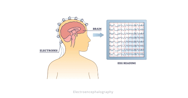
- Electroencephalography (EEG) is a medical test which measures brain electrical activity generated by neurons.
- It is a non-invasive neuroimaging technique that provides valuable insights into neural function and dysfunction.
- It is simple, affordable, portable, and safe compared to other methods like MRI.
- EEG excels at capturing rapid changes in brain activity, making it an invaluable tool for epilepsy diagnosis and monitoring.
- However, its limitations to mainly detects surface brain signals and has difficulty pinpointing activity origins.
- This versatile technique has far-reaching applications in diagnosing neurological disorders, monitoring anesthesia effects, studying sleep patterns, and confirming brain death.
Background
- Richard Caton first noticed electrical signals in animal brains in 1875.
- Adolf Beck and Vladimir Pravdich-Neminsky expanded on Caton’s work, with Pravdich-Neminsky recording the first EEG of a dog.
- Hans Berger recorded the first human EEG in 1924.
How does it work?
- It tracks neurons’ movement of charged particles, helping doctors diagnose epilepsy, monitor anesthesia, study sleep patterns, and confirm brain death.
- Neurons’ interactions create waves of electrical activity detected by scalp electrodes.
- Setting up an EEG involves applying gel and placing electrodes accurately, which can be affected by thick hair.
Latest News
- 17 July 2024:
- This year marks the 100th anniversary of the first human electroencephalography (EEG) by German physiologist Hans Berger.


Responses